Keynote Speakers
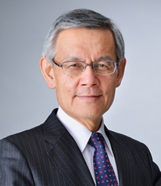
Dr. Nagara Tamaki
Professor, Department of Radiology, Graduate School of Medical Science, Kyoto Prefectural University of Medicine, JapanSpeech Title: Molecular Imaging by PET
Abstract: Recent progresses in imaging permits various non-invasive molecular imaging in vivo. Among them, positron emission tomography (PET) has recently been applied for quantitative molecular imaging using various molecular probes for human studies. F-18 labeled fluorodeoxyglucose (FDG) has been commonly used for assessing glucose uptake and metabolism in vivo using PET system.
FDG-PET permits detection and staging of malignant tumors. Thus, this technique is valuable for selecting optimal treatment planning (Precision Medicine). It has recently been used for treatment monitoring and predicting outcome with use of quantitative assessment of FDG uptake in malignant tumors. Furthermore, it holds a new clinical value for suitable radiation planning of malignant tumors.
Most recently, PET has been used in the field of neurology and cardiology as well. PET has a potential for predicting early stage of Alzheimer’s disease. In addition, FDG-PET has a new role for identifying active lesions in cardiovascular diseases.
This keynote lecture will cover advantages of molecular imaging by PET and introduction of various clinical applications on oncology, neurology, and cardiovascular fields.
Biography: After graduate from Medical School in Kyoto University in 1978, I have specialized Nuclear Medicine and Molecular Imaging for over 40 years with use of various nuclear medicine radiopharmaceuticals and SPECT/PET system. We have published several key scientific papers in the field of nuclear cardiology during my PhD course. In 1995, I have promoted as professor and director, department of nuclear medicine, Hokkaido University. Since then, I have focused on new clinical and basic studies using PET and molecular imaging. We have published over 500 original articles and over 60 books and chapters in this field. In addition, I have spent a lot of efforts for educating students and young fellows in our field.
I have had three times in studying abroad. I studied in Kansas City, Missouri, USA as an exchange student in high school in 1970-1971. Also, I stayed in Massachusetts General Hospital and Harvard Medical School as a research fellow in 1984-1986. I worked on renal blood flow measurement in animal models using PET. In addition, I had a chance to apply new portable cardiac function monitor in various cardiac patients. I have published several key papers in these fields. More recently, I visited Munchen Technical University, Germany as a visiting professor for three months in 2002. These chances have helped me for making wonderful friends all over the world, particularly in the field of nuclear medicine and molecular imaging.
After receiving degree of Emeritus Professor in Hokkaido University, I have moved to Kyoto Prefectural University of Medicine, as a professor in Department of Radiology in 2017. We are managing oncology PET center with in-house cyclotron in this University.
I have received (1) Japanese Society of Nuclear Medicine Award in 1990, (2) Georg de Hevesy Nuclear Medicine Pioneer Award in Society of Nuclear Medicine (SNM) in 1998, (3) SNM Cardiovascular Council, Hermann Blumgart Award in 2009, and (4) Award from Hokkaido Medical Association and Governor of Hokkaido in 2013.
Dr. Simon James Fong
Data Analytics and Collaborative Computing LaboratoryAssociate Professor, University of Macau, Macau SAR, China
Honorary Professor, Durban University of Technology, Durban, South Africa
Adjunct Professor, ZIAT of Chinese Academy of Sciences, Zhuhai, China
Adjunct Professor, Xi'an Polytechnic University, Xi'an, China
Senior Visiting Scholar, Tsinghua University, Beijing, China
Speech Title: AI in Histopathological Image Analysis for Cancer Detection: Past, Present and Future
Abstract: Since the inception of virtual microscopy in the 1990's, histology image analysis has migrated from optical microscope to visual inspection over digital histological images that were scanned and generated at high resolution. Detection of cancer and estimation of its prognosis are tedious process from the digital whole slide images because of its sheer area and complexity. Recently, by the efforts of multi-disciplinary research, artificial intelligence methods especially computer vision, object recognition, deep learning and XAI have become popular aids to grading and staging of cancer diagnoses and prognosis, by automatically analyzing over digital histopathology images. It was claimed that AI has helped pathologists to lower down the assessment errors by magnitude. Most of the research literature reports about identifying the cell types, segmenting the tumour and the cells. Current trend has it that the recognition of cells is done by deep learning due to its effectiveness in learning the features of the cells and recognizing them by their features. However, the accuracy and certainty of those histological deep learning methods are bottle-necked at the image level. Recognition rate is never perfect from solely the images. Lately it is discovered that the tumour immune microenvironment (TME) consists of many heterogeneous cell types that engage in extensive crosstalk among the cancer, immune and stroma tissues. In addition to just the cell types, the spatial organization of these different cell types in TME are observed as biomarkers for predicting metastasis, prognosis and drug responses via scRNAseq and spatial transcriptomics technologies. It opens up a new arena where AI can extend its power in analyzing the localization of cells, types of cells and their interactions, as a whole for further improving the prediction accuracy. In this talk, we will narrate the brief history, the current practices and future prospect of applications of AI on histological image analysis. This speech puts the future of AI research over histological images into a perspective that extra dimensions of analysis are in need for inferring more insights from histological image analysis. Novel models in this perspective are introduced. Future opportunities by integrating digital histopathology images with molecular omic data, spatial information, as well as meta-level analysis will be discussed for better understanding of a tumour ecosystem. During the keynote speech, our lab principal researcher, Dr. Gloria Li, will demonstrate several deep learning applications which are the cornerstone AI techniques over histopathological image analysis.
Biography: Simon Fong graduated from La Trobe University, Australia, with a 1st Class Honour BEng. Computer Systems degree and a PhD. Computer Science degree in 1993 and 1998 respectively. Simon is now working as an Associate Professor at the Computer and Information Science Department of the University of Macau. He is a co-founder of the Data Analytics and Collaborative Computing Research Group in the Faculty of Science and Technology. Prior to his academic career, Simon took up various managerial and technical posts, such as systems engineer, IT consultant and e-commerce director in Australia and Asia. Dr. Fong has published over 500 international conference and peer-reviewed journal papers, mostly in the areas of data mining, data stream mining, e-health, meta-heuristics optimization algorithms, and their applications. He serves on the editorial boards of the Journal of Network and Computer Applications of Elsevier, IEEE IT Professional Magazine, and various special issues of SCIE-indexed journals. Simon is also an active researcher with leading positions such as Vice-chair of IEEE Computational Intelligence Society (CIS) Task Force on "Business Intelligence & Knowledge Management", and Vice-director of International Consortium for Optimization and Modelling in Science and Industry (iCOMSI). In 2021, Dr. Fong was awarded with a scholar title of Overseas High-Level Talent Recruitment Programs.
List of speakers will be updated soon...


